Surveillance of zoonoses in livestock and humans: experiences from AHITI interns cohort 5

Surveillance of zoonoses in livestock and humans: experiences from AHITI interns cohort 5

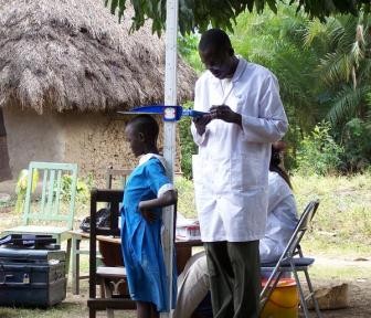
Our participation in the ZooLinK suite of projects will remain memorable. We have acquired sufficient knowledge and experience through the exposure given to us by ZooLinK staff and our participation in the target areas of the project. Since we joined the project on May 2018, we have rotated among the three functional units of the project, namely: (1) veterinary team who visit the livestock markets and slaughterhouses; (2) laboratory team and (3) clinicians team who visit the health centres. The following report will focus on the veterinary team. It describes the activities carried out therein and their relevance to the project.

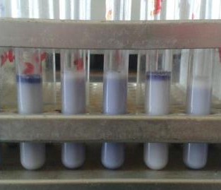
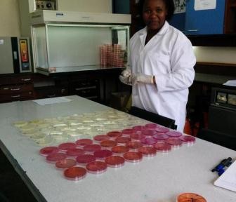
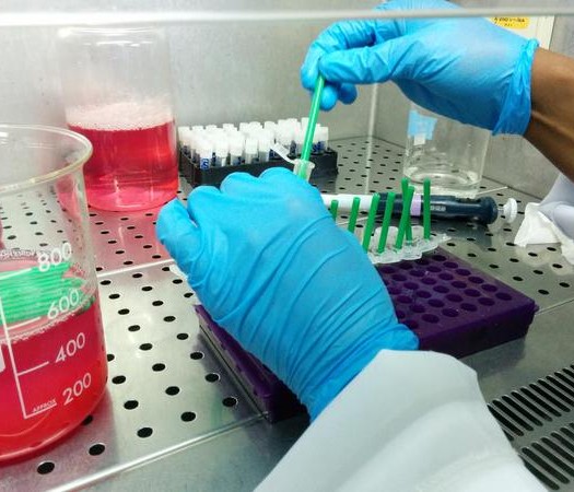
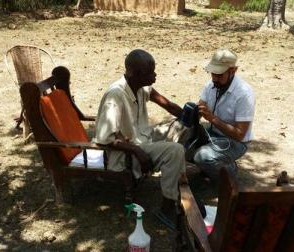
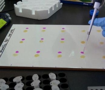
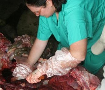
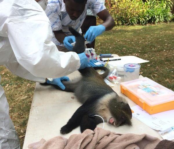
Two of the interns working in the laboratory (foreground)

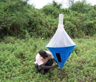
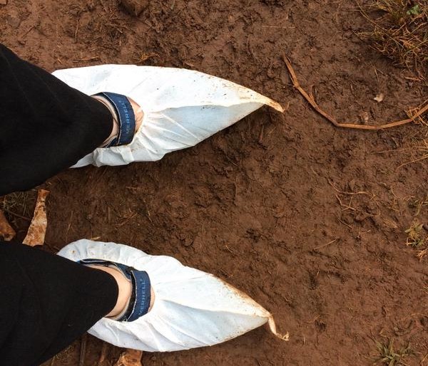
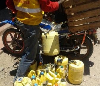
A normal ZooLinK day begins with packing the field car with the required consumables a day before the field. Such consumables include; red and purple topped vacutainers, nasal swabs, digital thermometer, heart girth measuring tape, ziplock bags, barcodes, consent forms, faecal pots, gloves, disinfectant, water, coveralls and gumboots etc.

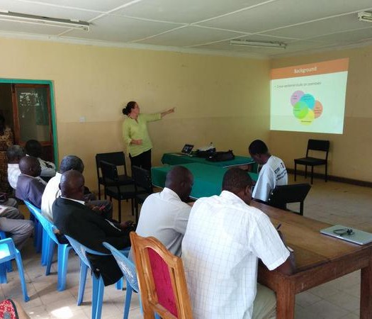
“…our internship has equipped us with adequate disease surveillance skills in the animal field that will help us to extend the knowledge of disease control to farmers…”

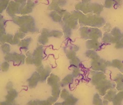
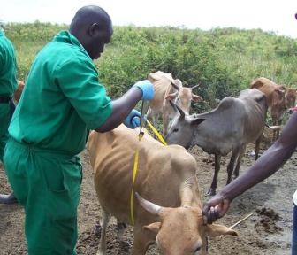
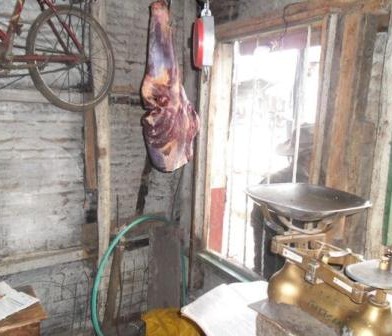
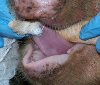
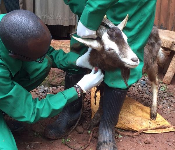
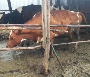
In the field, the veterinary team splits into two groups; one group works at the livestock markets and the other at the slaughterhouse. Upon arrival, at the livestock market, the animal is randomly selected and the owner identified to seek consent for sampling the animal and to answer a few questions. If he/she agrees, he/she signs two consent forms one of which goes with the animal owner while the other one remains for ZooLinK records. Before sampling, the animal is humanely restrained to ensure the safety of the animal, handler and person collecting the samples. Physical examination begins before the actual sample collection. Which entails checking for any abnormal discharges from the mouth, eyes, genitals and nose. On the skin swellings and injuries are recorded when present. Nature of the ocular mucous membranes is assessed and recorded, the mouth is checked for any lesions and sores as well the ageing is done from the dentition. The pre-scapula lymph nodes are palpated on both sides to ascertain any enlargement. Lifting of the loose skin of the neck is done to test for skin elasticity. The body condition of the animal is cored in a scale of 1-5. The fleece condition is recorded as either rough or normal and a tape measure used to measure the heart-girth to estimate the weight of the animal. The temperature is taken per-rectal. After the physical examination, the actual collection of the samples begins. Blood is collected from the jugular vein into a red top vacutainer (plain blood) for serology and an EDTA-purple top vacutainer (uncoagulated blood) for parasitology and hematology.

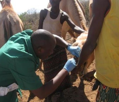
One of the AHITI interns sampling blood from a sheep

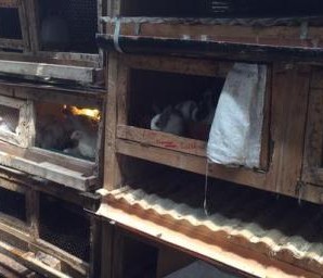
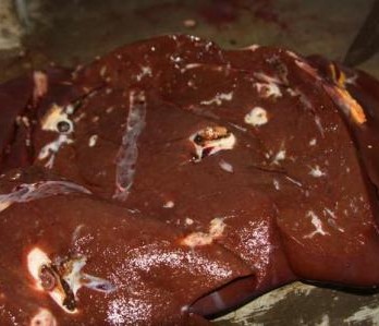
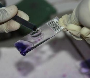
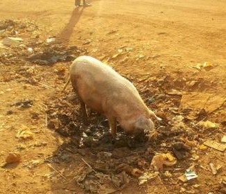
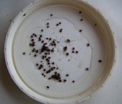
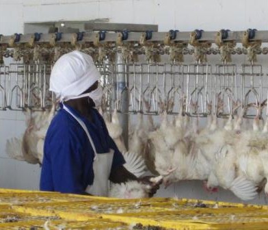
Nasal swabs are used to collect swabs from the nose. Nasal swabs are later cultured in the lab and used to test for the presence of Staphylococcus aureus. Fresh faeces are collected per-rectal and placed into a faecal pot. The faecal sample is cultured in the lab to determine the presence of E. coli, Salmonella and Campylobacter. External parasites like ticks, lice etc. are also collected if encountered. The same procedure takes place in the slaughterhouses but in addition, post-motem lesions like cysts, flukes, are recorded and collected inclusive of mesenteric lymph nodes from the pigs.

We are glad to declare that our internship has equipped us with adequate disease surveillance skills in the animal field that will help us to extend the knowledge of disease control to farmers and other stakeholders back at home.

This article was authored by the cohort 5 interns from the Animal Health and Industry Training Institute (AHITI): Sarah Nyambura, John Parkasio and Silas Muriithi.

Establishing a serum bank of samples from confirmed cysticercosis positive and negative pigs

Establishing a serum bank of samples from confirmed cysticercosis positive and negative pigs

This serum bank will serve as a platform for future development and validation of diagnostic tools that will allow for a quicker and more accurate diagnosis of porcine cysticercosis. The disease is zoonotic, meaning that it can be transmitted between humans and animals (pigs). The tapeworm, Taenia solium, causes taeniasis in people and can cause abdominal pain, diarrhoea, nausea and indigestion. The larval stage of the worm can infect both pigs and people. In people, the larval stage can become encysted in the brain and/or spinal cord, causing neuro-cysticercosis. This is an important cause of acquired epilepsy – a debilitating disease. The signs of the disease in humans include seizures, chronic headaches, dementia, and may result in death.

“The project aims to establish a bank of serum samples from confirmed cysticercosis positive and negative pigs.”

Fig.1. Making 3mm thick slices

We have organized to visit and buy pigs from 13 slaughter slabs spread across Busia and Kakamega Counties. The process involves contacting a trader/farmer at the slaughterhouse to deliver a pig on site. On the day of slaughter, intricate bargaining with the trader/farmer to ensure value for money ensues. This is a complex process given that the pricing is usually fluid, with no clear parameters to determine the price. The prices are usually based on the physical appearance of the pig which requires a lot of experience. Once the prices have been settled, photos of the pig are taken, and demographic information, such as age, heart-girth measurement and back length, are recorded. The blood is collected at ante-mortem and lingual palpation is performed. The pig is slaughtered and weighted perimortem, and then skinned. This is a source of amusement among the butchers who have christened this ‘naked pig carcass’ as Mbuzi ulaya loosely translating to a ‘European goat.’ The carcass, together with the head, lungs, liver and diaphragm, are chilled overnight and sliced (Fig.1.) in the morning.

The slices ought to be at least 3mm thick to ensure any cyst present can be exposed. This is a laborious process that usually takes 3-4 hours to complete. The most recent studies carried out in the same region recorded a prevalence of 37.6% using a serological method, and 34.4% by lingual palpation. It is such findings, combined with an increase in pig keeping and consumption, that call for such a study. Currently, there exist several serological tests which detect circulating T. solium cyst antigens in humans and animals. Yet most of these tests have poor specificity, leading to a large number of false positives and hence, limiting their diagnostic capacity. We look forward to sharing more insights from this project in subsequent newsletters.

This article was authored by Dr Maurice Karani who is ZooLinK Research Assistant and Field Coordinator.

Zoonoses in Livestock in Kenya – The Beginnings of Surveillance

Zoonoses in Livestock in Kenya – The Beginnings of Surveillance

By Steven Kemp, PhD student, University of Liverpool

After a period of intense lab work at both KEMRI and the UK, investigating the patterns of antimicrobial resistance in faecal bacteria isolated from slaughterhouse workers in Busia County and the surrounding areas, I have returned to Kenya to begin the next phase of my PhD project.

ZooLinK is a cyclical programme which aims to set up surveillance systems of both human and animal health sectors over a long period of time. Surveillance of disease is particularly important, as the more information we have, the better we can treat diseases in both human and animal sectors. Recent research by colleagues indicates that the incidence of several zoonotic diseases, including E. coli, Salmonella sp. and others are vastly underestimated.

In recent times, we often hear about how we should now look to conform to the ‘One Health’ approach; this is where, in order to combat issues surrounding antimicrobial resistance and associated issues effectively, intersectoral approaches which share the cost and responsibility evenly between environmental, human & veterinary health professionals is required. In theory, this would be a perfect way to help educate and better promote antimicrobial stewardship.

Currently, I have large amounts of data on access to, use of, and perceptions of antimicrobials from a variety of parties, including animal healthcare workers, district veterinary offices, farmers and agrovet shops. Over the last three months, I have added to this repository by investigating the amounts of antibiotic resistance found in E. coli, which have been isolated from the faeces of workers in 142 slaughterhouses which were selected in western Kenya. These included slaughterhouses in Busia County and the surrounding Kakamega and Bungoma counties.

For the next portion of this study, I am attempting to collect four different sets of samples – to complete the ‘picture’. I will attempt to collect both human and animal faecal samples, from farmers and farm animals, water samples (to determine if there are patterns of resistance in animals which share common grazing grounds) and environmental samples (from the inside of homesteads, where animals are allowed to roam). By covering all of these bases, we will be able to eventually determine not only if there is transfer of antimicrobial resistance between animals and humans and the environment, but also which direction it is going in.

Typical small-holder farm in Funyula, Busia. Most farmers manage between 5-25 cattle.

Example of environment which may also be a good idea to sample in the future. If antimicrobial resistance can be found in the envi-ronment, then why not in wild animals such as these Zebra?

Non-typhoidal Salmonella (NTS) in pigs in Busia, Nairobi and Malawi

Non-typhoidal Salmonella (NTS) in pigs in Busia, Nairobi and Malawi

This blog post was authored by Catherine Wilson an MRES Student from the University of Liverpool attached under our #ZooLink project

I am investigating the prevalence of Non-typhoidal Salmonella (NTS) in pigs in both Kenya and Malawi in extensive, low input production systems.  The aim is to determine whether invasive NTS are present in the pig population of three study areas; one rural and one urban area in Kenya and one rural region of Malawi. In sub-Saharan Africa, NTS is a leading cause of human mortality, particularly in the very young, old, malnourished, or those suffering from co-morbidities such as HIV or malaria.

Pig slaughter slab in Bumala

Pig slaughter slab in Bumala

An invasive NTS serovar has been found to be able to cause severe disease in chickens; suspicion is therefore arising that transmission between humans is not the sole route of spread of NTS, and that zoonotic transmission, especially from pigs, may have a role to play in the epidemiology of the disease. Should this invasive strain of bacteria be found in pigs, we will assess whether the same serovar clinically affects humans in the same geographical location, using data already gathered from human hospitals. A correlation between the two would indicate that zoonotic transmission may be occurring.

The final part of this study will assess the presence of drug resistance in the strains of NTS isolated from pigs, and whether this bears any correlation to a similar antimicrobial resistance pattern of NTS to that previously detected in humans in the same area.  Should antimicrobial resistance be detected, other management techniques for the swine, such alterations in husbandry and hygiene, may be trialed.  In the longer-term vaccination development may be a possibility as an important method of preventing zoonotic disease transmission in the study areas, for which research is currently in the very early stages.

For sampling,  both faecal and mesenteric lymph nodes samples were collected post mortem from 256 pigs in Busia and 304 pigs in Nairobi.  The location in which the pigs were reared, as well as details of signalment, any previous antibiotic treatment if known and the method of transport of the pig to the slaughterhouse, were recorded for each individual pig.

Samples were processed at the Busia Field Lab and ILRI laboratories respectively. Culture and serotyping was carried out to confirm the presence of Salmonella followed by antimicrobial susceptibility testing to a range of antibiotics.  Positive isolates have then been stored for transport to the UK, where whole genome sequencing will be undertaken to identify the presence of any antimicrobial resistance genes. Once the results have returned, analysis is planned compare antimicrobial resistance profiles of the pig samples to those of humans in the same geographical location, to assess whether zoonotic transmission may be occurring.

Laboratory capacity to diagnose Mycobacterium bovis in East Africa

Laboratory capacity to diagnose Mycobacterium bovis in East Africa

The full report can be accessed at this link: http://www.rr-africa.oie.int/docspdf/en/2016/CHEROTICH1.pdf

A report by Dr. Chepkwony submitted to the OIE- Regional Representation for Africa explores the diagnostic capacities at different scales for both human and animal national tuberculosis reference laboratories in Kenya, Uganda and Tanzania to diagnose Mycobacterium bovis. One recommendation put forward is that national governments should invest in new and more accurate diagnostic technologies for detecting zoonotic tuberculosis. Moreover, it is important to utilize regional and international partnerships and carefully determine how to link these new tests and incorporate them within a country’s national diagnostic algorithm.

Pin It on Pinterest