Urban Zoo Team — Out and About

Urban Zoo Team — Out and About

The International Symposium for Veterinary Epidemiology and Economics (ISVEE) 14th conference was held in Merida, Yucatan in Mexico from 3rd to 7th November 2015. The Urban Zoo group was well represented by Patrick Muinde, Pablo Alarcon, Paula Dominguez, Maud Carron, Joshua Onono, Judy Bettridge, Annie Cook and Jonathan Rushton.

Out and about

The theme of the conference was “Veterinary Epidemiology & Economics: Planning our future” and this group gave oral presentations in several streams in the confer-ence, including presenting much of the work on value chains; but also gave talks on food nutrition, zoonoses, animal health economics, statistics and food safety.

In addition to having some excellent talks, the social side of the conference was also highly enjoyable, with many opportunities to sample local food and see some of the historical sites around Yucatan. This also provided a chance to meet and discuss science with colleagues from around the globe.

Co PI’s Letter: Microbiology Thread

Co PI’s Letter: Microbiology Thread

On a dark and cold November afternoon, Edinburgh feels very far from Nairobi and the UrbanZoo project. But I like to think that we still have a useful contribution to make, not least by helping to put together different pieces of research together to reveal the “big picture”. It is going to be a very unusual picture though; we want to construct a kind of map of Nairobi from the point of view of a ubiquitous bacterium, Escherichia coli. We want to find out where different strains of E. coli live and how they move through the different compartments that make up Nairobi: healthy people  in the community, sick children in clinics, wildlife, food, water and waste and especially the livestock species that live in the city.

Those urban livestock are central to UrbanZoo: we want to know how they fit into the ecology of E. coli in Nairobi and, in doing so, we hope to illuminate a general theme of the entire ESEI programme (of which UrbanZoo is one of 3 funded projects). We will be testing the idea that urban livestock are a risk factor for outbreaks of emerging diseases (not just E. coli-related disease) in cities everywhere.

Though often stated, that hypothesis has rarely been tested. It might not even be true: we shall see. It all starts with the highly structured collection of samples by the field teams, who are systematically working their way around Nairobi as they conduct the 99 Household survey. The microbiology teams will then give us a first glimpse of the big picture by carefully typing the E. coli isolated from those samples.

That work is vital, but although standard typing methods can tell us whether two samples share similar kinds of E. coli, they often cannot tell us the direction of movement. For example, if samples from a child and a pig in the same household contain very similar E. coli, does it mean that the pig infected the child, the child infected the pig, or they were both infected from some other source? The state-of-the-art tool for answering those questions is kind of statistical analysis called phylodynamics. Our phylodynamic analyses will use two kinds of information: whole genome sequences of UrbanZoo E. coli; and the metadata – place, time and host
species in particular – that go with those sequences.

That is why everyone is working so hard to obtain the best possible set of isolates for sequencing and all the field and typing data that go with them, why we are spending so much money on whole genome sequencing, and why Melissa Ward is devoting so
much of her time to doing the phylodynamics analyses.

Over the next 12 months or so we hope to build up a collection of E. coli sequences that is by far the best of its kind in the world and that gives us completely new insights into the ecology of the bacterium within the extremely complex ecosystem that is Nairobi. We hope to reveal a picture that no one else has ever seen before. I can’t wait.

Mark Woolhouse is Professor of Infectious Disease Epidemiology at the University of Edinburgh in Scotland.

Studying African camels is key to learning more about the MERS virus

Overview

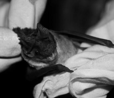
African camels could hold important clues to controlling the potential spread of a respiratory disease transmitted by the animals.

For many years African camels have lived with the disease and the risk of it spreading to humans is still low. But more research is necessary to understand the disease better. This is even more important given the confirmation that the chains of transmission of the human Middle East Respiratory Syndrome Coronavirus (MERS-CoV) infection originated from contact with camels. MERS was first recognised in 2012.

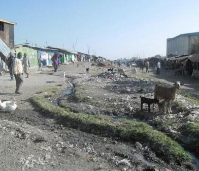
Camels are an extremely important source of livelihood, nutrition and income in Africa. They are especially common in arid and semi-arid areas of the continent, particularly in East Africa. But having these animals around may not be risk-free for humans.

However there have been no human case of MERS diagnosed in Africa. This could be because of limited clinical or epidemiological surveillance for the virus where infections have gone unrecognised. It could also be because there is simply no zoonotic, human infective virus circulating in sub-Saharan Africa, or indeed because risk factors for transmission differ in the two regions.

MERS from an African perspective

Looking at Africa, what do we know about the disease and the potential risk of transmission?

The disease usually affects patients who are in some way immune-compromised or suffering from other conditions like diabetes, lung and liver disease.

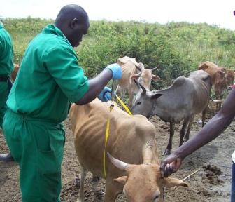
Livestock dependent people in the Horn of Africa region who suffer from malnutrition are potentially at risk of contracting the disease. The case fatality rate of MERS is high, at around 37%. Outbreaks in other parts of the world like South Korea have been linked to individuals originally acquiring infections from chains of transmission originating in the Middle East and travelling.

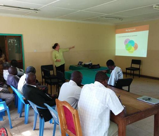
In October the Centre for Pastoral Areas and Livestock Development at the Intergovernmental Authority on Development and the UN Food and Agriculture Organisation co-hosted a scientific and policy meeting to discuss the MERS virus. The aim was to improve the understanding of this pathogen and its implications to the Horn of Africa region.

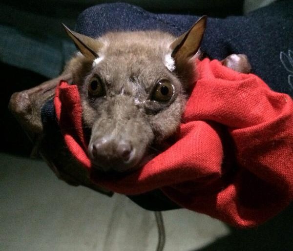
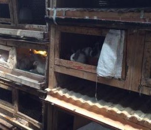
The meeting was prompted by the likely role of the dromedary camel as a reservoir of infection for MERS-CoV, and the high density of and trade importance of camels in the Horn of Africa region. The region supports more than 60% of the world’s population of single humped camels.

There are two types of camels worldwide. The dromedary camel is found in Africa and the Middle East; and the Bactrian Camel, found in Asia.
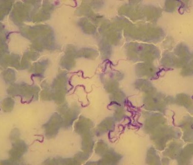
The virus in camels

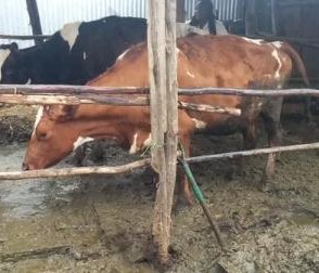
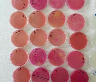
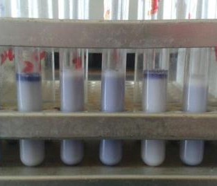
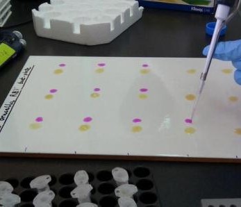
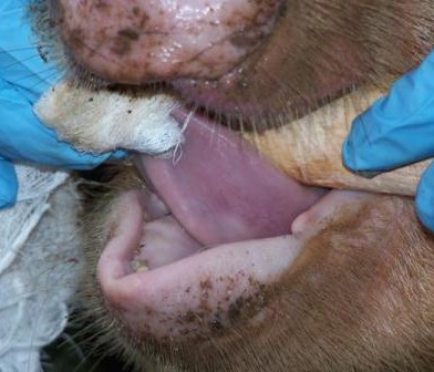
Studies in Kenya and elsewhere show that, despite its recent identification as a human pathogen, MERS has been circulating for many years over wide geographical areas. Camel sera collected as far back as 1983 shows high rates of seroconversion to the virus. This means that the animals have been infected, probably by a transient respiratory disease, and recovered.

MERS in camels, it seems, is much like being infected by the common cold. Some populations of camels in Kenya (which has the third largest camel population in East Africa) tested recently show seroconversion rates of 47%. This is a widespread virus that is actively circulating and has been around for a long time.

A crucial question in understanding the disease is establishing what the human risk is when the virus circulates so freely in the reservoir host. It is vital to learn whether dromedary camels in Africa harbour the same MERS-CoV as detected in the Arabian Peninsula. If so, or if not, is the epidemiology of the virus similar?

This is despite the high seroconversion rates, as the virus appears to affect camels early in life – possibly before they have weaned – and self cure within a matter of weeks.

Sampling a camel_Eric_Fevre

Mapping trade routes and understanding the population structure of African camels better with their population density will also be key foresight information should large scale disease control interventions ever be necessary.

More research is key

The appearance of a new disease in a widely distributed reservoir host is a worrying prospect. It does however seem that camels in Africa have been living with MERS-CoV for a long time. While the risk of spill over to humans from this population cannot yet be discounted, it appears to be, for now, a low risk.

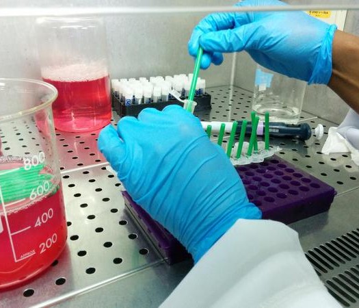
Even though the Middle East has seen outbreaks of a virus with zoonotic potential, it might be that the mutations required to make this possible have only evolved recently and in that locality. The newly acquired zoonotic potential may not be widespread. To better understand if this is the case, active efforts are underway in sub-Saharan Africa to isolate the virus itself and genetically type it.

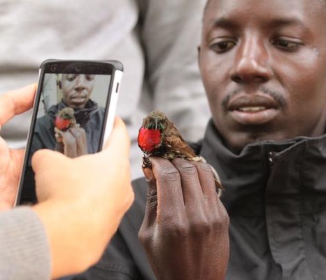
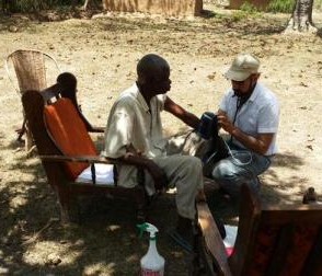
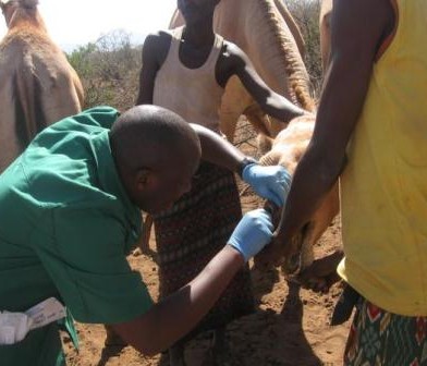
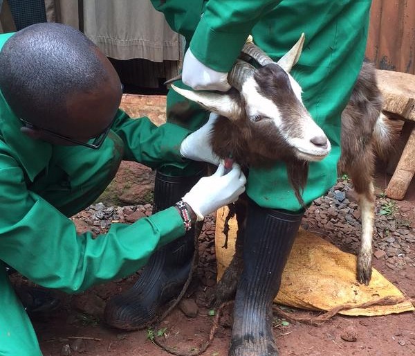
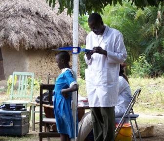
In conjunction with genetic studies of the virus, work is also underway to determine whether people, and particularly those at high potential risk such as camel herders and slaughterhouse workers, have also seroconverted to the virus. This would demonstrate that human infections have taken place.

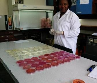
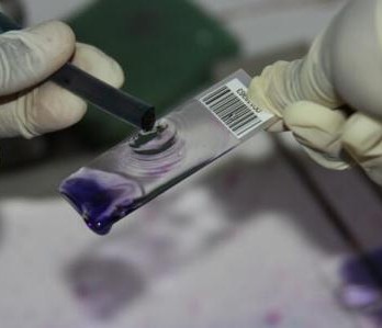
Part of the effort to do this involves building local diagnostic capacity in Africa, to ensure that such at-risk populations can be monitored through time and increase the speed of a public health response if required. As with many diseases, a diagnostic test that could be used in the field would be ideal for such monitoring.

For long term preparation, a key research priority is to understand the continental distribution and diversity of camel populations themselves. The camel is very much under-researched, compared to other livestock such as cattle, goats and sheep, despite its importance to rural livelihoods in many areas.

This gives the livestock and health communities the opportunity to study and better understand this virus, ideally working on a joint agenda that shares knowledge. An example of this is the One Health philosophy between sectors that benefits all.

Dr Joerg Jores of the International Livestock Research Institute featured as a co-author on the piece.


This article was authored by Eric Fevre (Professor of Veterinary Infectious Diseases, University of Liverpool) and  originally published on The Conversation. Read the original article by Clicking here

Conversation logo


99 HouseHold Study: Sykes monkey

Identification

It is known as Kima/Nchima in Swahili and having other names such as blue monkeys or samango’s or white-throated monkey. The majority have a grey coloration, but they do have a blackish tail, limbs and shoulders with some chestnutty patches on their back and face. Finally they have a white chin and throat and a white ruff that extends around part of their neck. They have gorgeous brown/orange eyes with a slightly large squashed nose.

Sykes monkeys have an average life span of around 27 years in the wild. They can range from 50 to 70cm in body length with the males weighing between 6-9kg and females 3-6kg.

Distribution

The Syke monkey is native to Angola; Burundi; Congo, The Democratic Republic of the; Ethiopia; Kenya; Malawi; Mozambique; Rwanda; Somalia; South Africa; South Sudan; Swaziland; Tanzania, United Republic of; Uganda; Zambia; Zimbabwe. They are semi-arboreal semi-terrestrial and are comfortable in both environments.

Habitat and Ecology

This species is present in many different forest types including lowland and montane tropical moist forest, riverine and gallery forest, delta forest and bamboo forest. Also found in sand forest. It can occur in secondary forest, logged forest and thickets. Group size ranges from 2 to more than 40 individuals. The gestation period is around 176 days after which a single young is usually born.

Diet

Their main diet is shoots, fruits, leaves, flowers and berries, but they do also consume eggs, insects and any opportunistic human food they can get their hands on (especially when it comes to unsuspecting tourists).

Behaviour

Sykes are very territorial and live in female bonded; single-male troops and the group size can vary between 4-65. The groups are very stable and peaceful with the resident male maintaining dominance for years and the sub-adult males being evicted readily. Sometimes an outsider male will learn to imitate the call of the group’s dominant male to attract the females away.

Reproduction

The mating system is polygynous, and there is a corresponding sexual dimorphism in size, with the males substantially the larger sex. Females normally give birth every two years, during the onset of the warm, rainy season; gestation is around five months, and the infants are born with fur and with their eyes open. Group sizes range from 10 to 40, containing only a single adult male.

IUCN classification

As a species Cercopithecus mitis are rated as ‘least concern’ by the IUCN as it is widespread, common, present in numerous protected areas, and there are no major threats. However, when considered at subspecies level their rating ranges from data deficient to critically endangered.

Reference
?

Examining the Sykes monkey

?

Blood sample collection from Sykes monkey

99 Household Study: Fruit bat

Description

The Fruit Bat is a family of several species of bats that are sometimes referred as Flying Foxes. They have a life expectancy of about 30 years. They vary in terms of their length ranging between below 6 to 40 centimeters and weighing about 1.6 kilograms. They have large eyes with an excellent vision and sense of smell that they use in finding food and for their own protection. Their sharp teeth and long tongue enable them to feed successfully on fruits.  The shape of their wings varies to adapt to the various sites and locations that they belong to and at times the different wing sizes and shapes has been attributed to their different feeding environments.

Anatomy and Behaviour

Fruit bats are relatively large bats. Their wings are long fingers covered by thin skin, and the body is furry. Males and females look alike. Like all bats, they have weak legs and don’t walk very well. Livingstone’s Fruit Bat (Pteropus livingstonii, from Africa) is one of the largest fruit bats; it has a wingspan of up to 6 ft (1.8 m). The long-tongued fruit bat (Macroglossus minimus) is one of the smallest fruit bats; it has a wingspan of 5-6 inches (13-15 cm).

The wings of the Fruit Bat is an adaptation to allow them to fly, stay warm when roosting. They belong to large colonies referred to as “camps” which is a safety mechanisms  and in every 8 females their can be 1 male. They feed on flower nectar and fruit juice.

Distribution and Habitat

The Fruit bats live in dense forests in Africa, Europe, Australia, and Asia. They tend to thrive well in forested and grassland areas with a lot of food especially fruit trees, especially in areas with warmer climates which propagate variety of trees to grow throughout the year. In certain times of the year they can travel long distances to find food. When roosting during the day, they do so high up in the trees which provides sufficient darkness that protects against various predators, or hide in crevices or other dark areas and typically live close to water bodies.

Reproduction

The gestation period of the Fruit Bat is about 6 months. After delivery the females nurture the young for a period of 6 weeks until the young is able to fly by itself.

Predators

Some of predators for the Fruit Bat are: Birds (e.g. hawk, eagle), snakes, cats.

Advantages to nature

As fruit bats fly from plant to plant getting food, they also pollinate the plants they visit. In addition, they disperse the plants’ seeds as they eat. Many plants, including some avocados, dates, mangos, and peaches, are dependent on these bats for either pollination or seed dispersal

Diseases transmission

Recent studies have demonstrated the potential of bats to act as reservoirs of zoonotic pathogens (as reviewed in Hayman et al.(2012). One example is the common and conspicuous straw-coloured fruit bat (Eidolon helvum), which has been identified by Banyard et al (2012) as a reservoir host for Lagos bat virus (LBV, family Rhabdoviridae, genus Lyssavirus) and henipaviruses (family Paramyxoviridae) by Hayman et al (2008) in mainland Africa.

?

Fruitbat (Epomophorus) during 99 Household study

Pin It on Pinterest